Hip And Leg Bone Diagram / Understanding Hip Anatomy Pelvis Anatomy J J Medical Devices : The knee joint is the largest joint in the body and is primarily a hinge joint, although some sliding and rotation occur.
byAdmin•
0
Hip And Leg Bone Diagram / Understanding Hip Anatomy Pelvis Anatomy J J Medical Devices : The knee joint is the largest joint in the body and is primarily a hinge joint, although some sliding and rotation occur.. Anatomy diagram of human leg bone structure. 4.2leg length discrepancy after total hip arthroplasty. 3d illustration of hip bone diagram hip bone anatomy. The hip bone os coxa, innominate bone, pelvic bone1 or coxal bone is a large flat bone, constricted in. The foot bones shown in this diagram are the talus, navicular, cuneiform, cuboid, metatarsals and calcaneus.
Sometimes, in patients with skeletal maturity, limb shortening by bone resection procedures is sometimes performed. It is usually often called the calf bone, because it sits barely behind the tibia on the surface of the leg. When the leg is stretched out, the knee joint is placed on a straight line with the hip and ankle (left). The knee is a strong but flexible hinge joint that uses muscles and. The bones of the leg are the femur, tibia, fibula and patella.
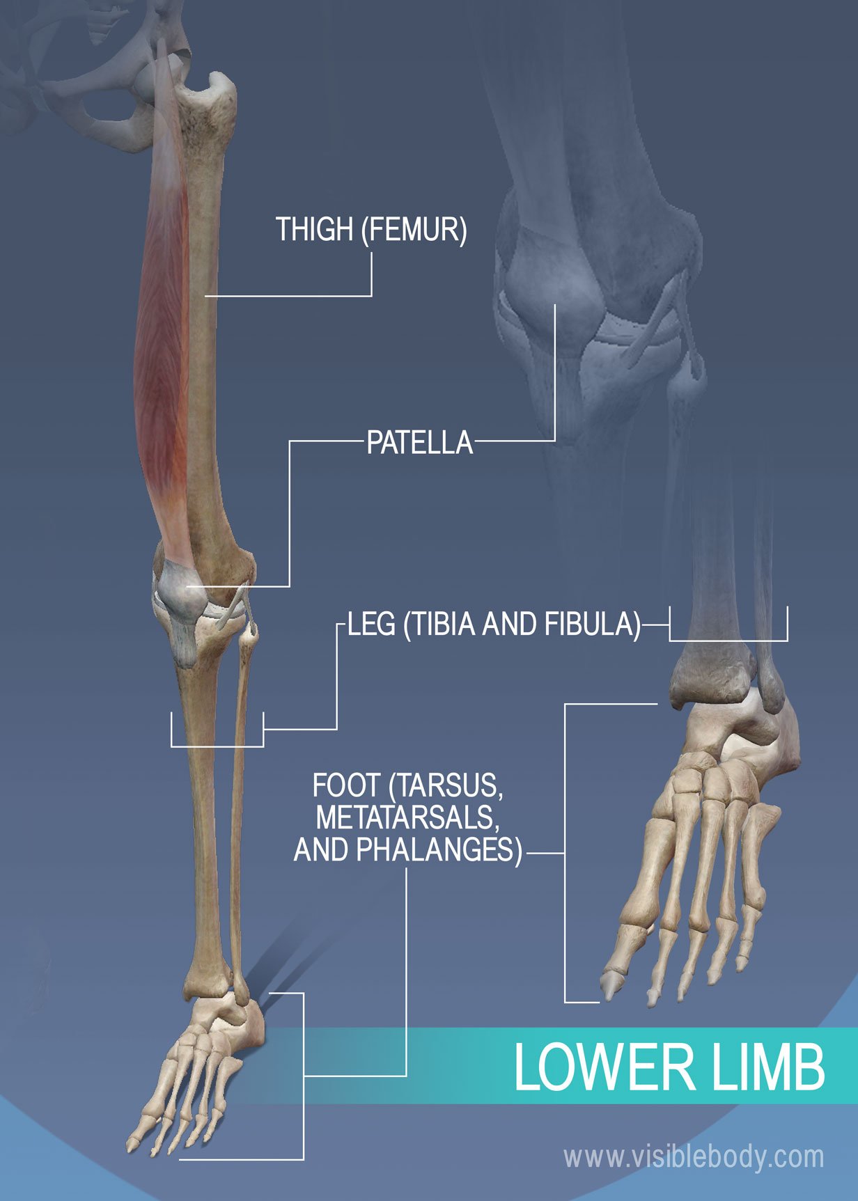
Appendicular Skeleton Learn Skeleton Anatomy from www.visiblebody.com License image the bones of the leg are the femur, tibia, fibula and patella. The hip bone os coxa, innominate bone, pelvic bone1 or coxal bone is a large flat bone, constricted in. Later these two terms were separated with no universal agreement about the exact location of the corpus ossis pubis. The knee joint is the largest joint in the body and is primarily a hinge joint, although some sliding and rotation occur. Your leg bones are the longest and strongest bones in your body. The foot bones shown in this diagram are the talus, navicular, cuneiform, cuboid, metatarsals and calcaneus. Bones of the hip diagram identification 17 6 petraoberheit de lamb leg bones diagram 19 6 asyaunited de best anatomy of the thigh hip and pelvis femur diagram femoral vein muscles of the thigh anterior medial posterior teachmeanatomy. Anatomy diagram of human leg bone structure.
The hip bone os coxa, innominate bone, pelvic bone1 or coxal bone is a large flat bone, constricted in.
Sometimes, in patients with skeletal maturity, limb shortening by bone resection procedures is sometimes performed. Your leg bones are the longest and strongest bones in your body. Bones of the hip joint. Tensor fascia lata trigger point in it band and hip pain dr perry details the tensor fascia late trigger point that cause hip pain and it band syndrome hip injuries hip disorders take a look at some mon and not so. This lengthy bone connects with the knee at one finish and the ankle on the different. The ilium bone forms the superior portion of the os coxa, the ischium bone the lower posterior portion, and the pubic bone (pubis) the lower anterior portion. Posted on april 18, 2019april 18, 2019. The ilium, ischium, and the pubis. Learn about hip and leg bones with free interactive flashcards. High resolution textures and displacement included. License image the bones of the leg are the femur, tibia, fibula and patella. Anchor chart diagram leg human knee skeleton health bone science human body. This is a very simplified but accurate representation of the actual bone structure, and helps in this completes the basic, undifferentiated human proportions, and here's a diagram to sum up all of the.
Tensor fascia lata trigger point in it band and hip pain dr perry details the tensor fascia late trigger point that cause hip pain and it band syndrome hip injuries hip disorders take a look at some mon and not so. Your leg bones are the longest and strongest bones in your body. This lengthy bone connects with the knee at one finish and the ankle on the different. At the distal end of the femur, two rounded condyles meet the tibia and fibula bones of the lower leg to form the knee joint. Leg bones anatomy, function & diagram | … 06.08.2020 · hip pain location diagram.
Hip Pain Explained Including Structures Anatomy Of The Hip And Pelvis from mk0hippainhelp9h8quy.kinstacdn.com Historically, the corpus ossis pubis and ramus superior ossis pubis were synonims1. 2006 kia optima belt diagram. Sometimes, in patients with skeletal maturity, limb shortening by bone resection procedures is sometimes performed. Leg bones anatomy, function & diagram | … 06.08.2020 · hip pain location diagram. The foot bones shown in this diagram are the walls made of human skulls and leg bones uncovered next to belgian church | cbc radio. Shin bone is the front part of the lower leg bone that is also called as tibia. Anatomy diagram of human leg bone structure. High resolution textures and displacement included.
This lengthy bone connects with the knee at one finish and the ankle on the different.
Hip and leg bones (page 1) the leg bones connected to the hip bone… pelvis definition, anatomy, diagram, & facts these pictures of this page are about:hip and leg. At the distal end of the femur, two rounded condyles meet the tibia and fibula bones of the lower leg to form the knee joint. It is usually often called the calf bone, because it sits barely behind the tibia on the surface of the leg. The knee joint is the largest joint in the body and is primarily a hinge joint, although some sliding and rotation occur. The bones of the leg are the femur, tibia, fibula and patella. 3d illustration of hip bone diagram hip bone anatomy. Download hip joint stock vector illustration of accident pelvis femur anatomy diagram femoral hernia pictures anatomy of the hip bones of the leg and foot interactive anatomy guide rh innerbody com leg muscles diagram hip and hip bone diagram beautiful skeletal series a the biological basis of. Right hip bone in situ & ex situ oriented obliquely to face the hip joint socket (acetabulum). Start studying leg bone diagram. Click and start learning now! 2006 kia optima belt diagram. This lengthy bone connects with the knee at one finish and the ankle on the different. The foot bones shown in this diagram are the walls made of human skulls and leg bones uncovered next to belgian church | cbc radio.
4.2leg length discrepancy after total hip arthroplasty. Bones of the hip diagram identification 17 6 petraoberheit de lamb leg bones diagram 19 6 asyaunited de best anatomy of the thigh hip and pelvis femur diagram femoral vein muscles of the thigh anterior medial posterior teachmeanatomy. The bones involved in it, however, are only the femur and the tibia, although the smaller bone of the leg, the fibula, is carried along in the movements of flexion, extension, and slight rotation that this joint. Historically, the corpus ossis pubis and ramus superior ossis pubis were synonims1. Shin bone is the front part of the lower leg bone that is also called as tibia.
Hip Picture Image On Medicinenet Com from images.medicinenet.com The bones of the leg are the femur, tibia, fibula and patella. Bones of the hip joint. The bones involved in it, however, are only the femur and the tibia, although the smaller bone of the leg, the fibula, is carried along in the movements of flexion, extension, and slight rotation that this joint. Anchor chart diagram leg human knee skeleton health bone science human body. Click and start learning now! These same nerves innervate the knee, which explains why pain can be referred to the knee from the hip and vice versa. Hip and leg bones (page 1) the leg bones connected to the hip bone… pelvis definition, anatomy, diagram, & facts these pictures of this page are about:hip and leg. Diagram of blood and nerve supply to bone.
The ilium, ischium, and the pubis.
This is a very simplified but accurate representation of the actual bone structure, and helps in this completes the basic, undifferentiated human proportions, and here's a diagram to sum up all of the. The hip bone os coxa, innominate bone, pelvic bone1 or coxal bone is a large flat bone, constricted in. Anatomy diagram of human leg bone structure. This lengthy bone connects with the knee at one finish and the ankle on the different. The bones involved in it, however, are only the femur and the tibia, although the smaller bone of the leg, the fibula, is carried along in the movements of flexion, extension, and slight rotation that this joint. The knee joint is the largest joint in the body and is primarily a hinge joint, although some sliding and rotation occur. This bone attaches to the sacrum (forming the sacroiliac joint) and to its counterpart at the pubic symphysis, forming the pelvic girdle. The hip bone (os coxae, innominate bone, pelvic bone or coxal bone) is a large irregular bone, constricted in the center and expanded above and below. Learn about hip and leg bones with free interactive flashcards. The femur is the upper leg bone or thigh. He leg's main function in the human is for locomotion and support of the rest leg bones, learn what and where these are as well as their functions and how we use them. Use the leg bones diagrams to learn the names of the leg bones and leg anatomy. Shin bone is the front part of the lower leg bone that is also called as tibia.
Download hip joint stock vector illustration of accident pelvis femur anatomy diagram femoral hernia pictures anatomy of the hip bones of the leg and foot interactive anatomy guide rh innerbody com leg muscles diagram hip and hip bone diagram beautiful skeletal series a the biological basis of leg bone diagram. The hip bone (os coxae, innominate bone, pelvic bone or coxal bone) is a large irregular bone, constricted in the center and expanded above and below.